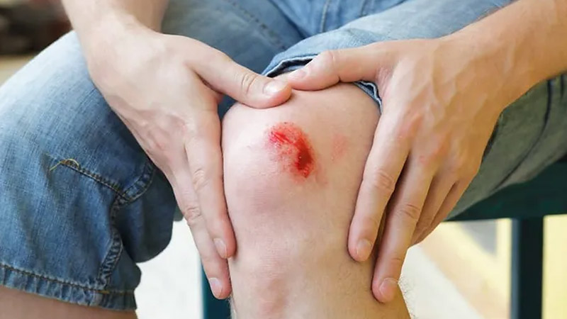
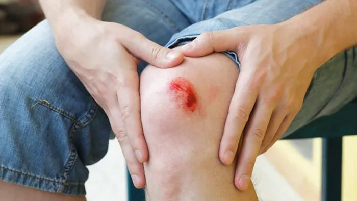
The skin is not impenetrable to injury, despite its powerful protection systems. It endures the cruel twists and turns of crazy luck every day and tries its best to keep us safe by recognizing and avoiding these threats.
A different defensive mechanism has been discovered by Elaine Fuchs’ team at Rockefeller University, and it doesn’t need an infection to kick in. It reacts to injury signals in damaged tissue, such as low oxygen levels from blood vessel rupture and scab development. The research is the first to identify a damage response mechanism that differs from yet runs concurrently with the traditional pathway activated by infections.
Interleukin-24 (IL24), whose gene is activated in skin epithelial stem cells near the wound edge, is at the center of the reaction. Once released, this secreted protein mobilizes a range of diverse cells to start the intricate healing process.
“Wound-edge epidermal stem cells primarily produce IL24, although numerous skin cells, including epithelial, fibroblastic, and endothelial cells, express the IL24 receptor and react to the signal. According to Fuchs, director of the Robin Chemers Neustein Laboratory of Mammalian Cell Biology and Development, IL24 “becomes an orchestrator that orchestrates tissue repair.”
Scientists have long known how the host defenses shield our bodies from hazards brought on by pathogens. But how does the body react to a wound that could or might not be caused by outside invaders? For instance, if we cut a finger while slicing a cucumber, we are aware of it right away since there is blood and pain. On a molecular level, it is still unclear how harm detection results in healing.
Type 1 interferons depend on the signaling molecules STAT1 and STAT2 to control the immune response to infections, but earlier studies by the Fuchs group had shown that a related transcription factor called STAT3 emerges during wound healing. Siqi Liu, who is a co-first author on both papers, aimed to determine where STAT3 initially appeared. A notable upstream cytokine that causes STAT3 activation in wounds is IL24.
The scientists used mice under germ-free settings in partnership with Daniel Mucida’s group at Rockefeller University and discovered that the wound-induced IL24 signaling cascade is unrelated to germs.
What damage signals, however, set off the cascade? The skin’s dermis, which contains capillaries and blood vessels, is often affected by wounds. Yun Ha Hur, a research assistant in the lab and a co-first author on the publication, adds, “We discovered that the epidermal stem cells sense the hypoxic environment of the wound.”
Epidermal stem cells are deprived of oxygen at the margin of the incision when blood vessels are broken and a scab develops. A positive feedback loop involving the transcription factors HIF1a and STAT3 amplifies the synthesis of IL24 near the wound edge as a result of this hypoxic condition, which serves as a warning sign for the health of the cells.
As a consequence, several cell types that express the IL24 receptor worked together to repair the wound by creating fibroblasts for new skin cells, replacing damaged epithelial cells, and mending capillaries. Together with Craig Thompson’s team at Memorial Sloan Kettering Cancer Center, the researchers demonstrated that altering oxygen levels might control Il24 gene expression.
After identifying epidermal stem cells as the source of the tissue-repair pathway, researchers examined the wound-healing process in mice that had their genes altered to render IL24 inactive. Without this essential protein, the healing process slowed down and took longer than it did in mice with healthy skin, lasting days longer.
They hypothesize that IL24 may be involved in the response to damage in other human organs with epithelial layers that serve as a protective coating. In epithelial lung tissue of patients with severe COVID-19 and in colonic tissue of individuals with ulcerative colitis, a chronic inflammatory bowel illness, higher IL24 activity has been detected in recent investigations.
It has become interesting to investigate IL24’s possible function in the damage response and disease pathology in these tissues in light of the finding of enhanced IL24 activity in other human organs with epithelial layers.
The elevated levels of IL24 activity in the epithelial lung tissue of patients with severe COVID-19 imply that the disease’s excessive inflammation and tissue destruction may be a result of this cytokine. This suggests that IL24 targeting may be a feasible treatment strategy to lessen the severity of COVID-19.
Similar to this, the higher IL24 activity seen in the colonic mucosa of ulcerative colitis patients raises the possibility that it contributes to the persistent inflammation and tissue damage that characterize this condition. Therefore, targeting IL24 may also be a potential therapeutic approach for the management of ulcerative colitis.
To completely comprehend the processes driving IL24 activity in these many tissues and illnesses, further investigation is required. However, the identification of its potential involvement in disease pathology and injury responses in numerous organs emphasizes the significance of this compound as a potential therapeutic target.


